
Understanding Upper Endoscopic Ultrasound
In the field of gastroenterology, the use of endoscopic ultrasound (EUS) has revolutionized the diagnosis and treatment of various gastrointestinal conditions. This minimally invasive procedure combines the power of endoscopy and ultrasound imaging to provide detailed visualization of the upper gastrointestinal (GI) tract and surrounding organs. Explore the intricacies of upper endoscopic ultrasound, its applications, the procedure itself, and what patients can expect before, during, and after the examination.
What is Upper Endoscopic Ultrasound?
Upper endoscopic ultrasound, also known as endoscopic sonography, is a specialized procedure that allows gastroenterologists to examine the upper GI tract, including the esophagus, stomach, and duodenum, using an endoscope equipped with an ultrasound probe. This combination of endoscopy and ultrasound technology enables detailed imaging of the GI tract and nearby structures, such as the pancreas, liver, and lymph nodes.
The Importance of Upper Endoscopic Ultrasound
Upper endoscopic ultrasound plays a crucial role in diagnosing and managing various gastrointestinal conditions. It allows for detecting and evaluating tumors, cysts, and other abnormalities within the upper GI tract and adjacent organs. It also aids in determining the stage of cancers and the extent of their spread, which helps guide treatment decisions. With its high-resolution imaging capabilities, EUS provides valuable insights into the structure and function of the upper digestive system, facilitating accurate diagnoses and tailored treatment plans.
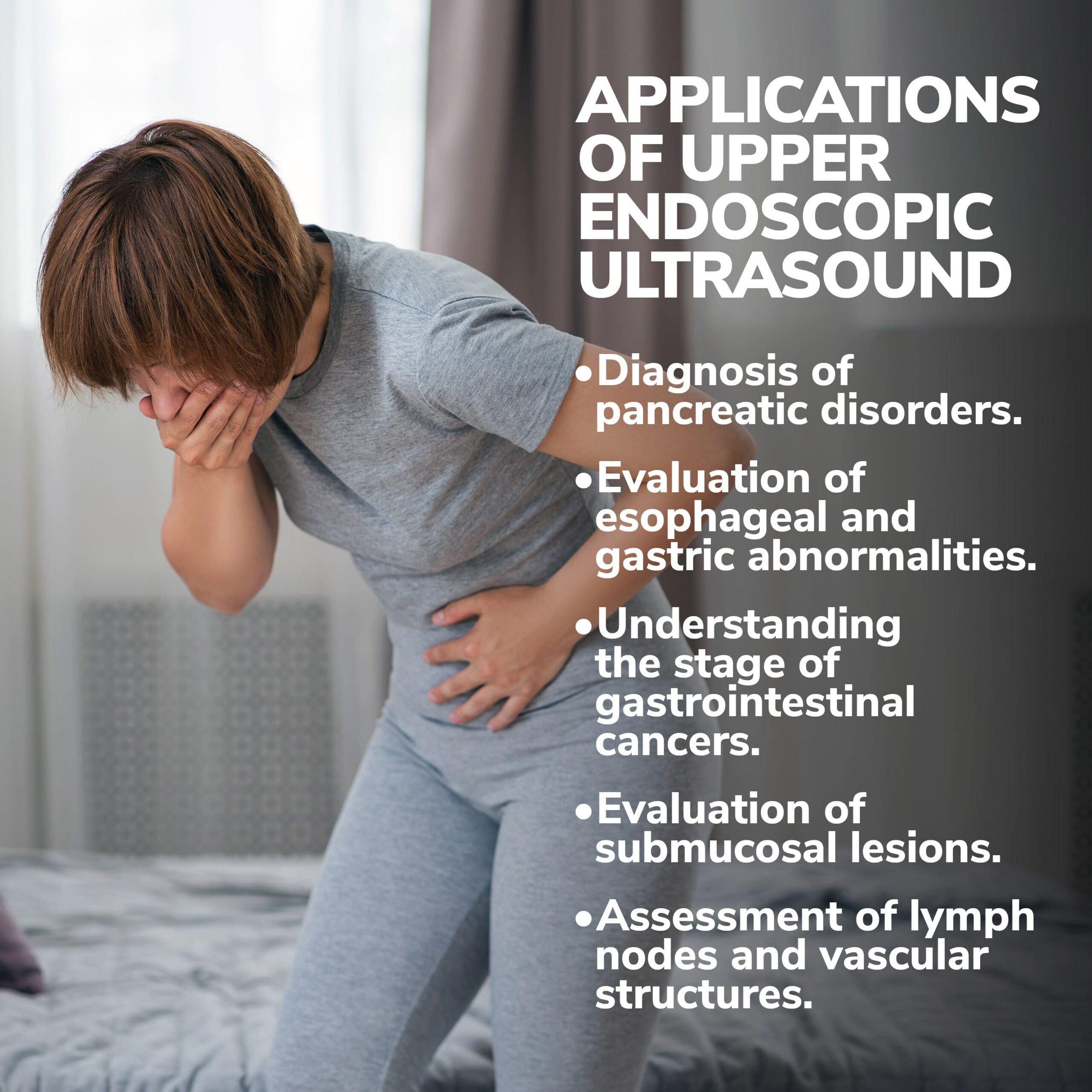
Applications of Upper Endoscopic Ultrasound
Upper endoscopic ultrasound offers a wide range of applications in gastroenterology. Here are some of the key uses of this procedure:
- Diagnosis of Pancreatic Disorders. One of the primary applications of upper endoscopic ultrasound is the evaluation of pancreatic disorders. EUS allows for detailed imaging of the pancreas, diagnosing conditions such as pancreatic cancer, pancreatitis, and pancreatic cysts. The ability to get tissue samples through a fine-needle aspiration (FNA) biopsy during the procedure further enhances diagnostic accuracy. This involves passing a thin needle through the endoscope to collect tissue samples for further analysis.
- Evaluation of Esophageal and Gastric Abnormalities. Upper endoscopic ultrasound is an invaluable tool for assessing esophageal and gastric abnormalities. It enables the detection of tumors, ulcers, and other lesions within the upper GI tract, providing essential information for diagnosis and treatment planning. EUS can also help evaluate the depth of tumor invasion, guiding decisions about surgical resection and other interventions.
- Understanding the Stage of Gastrointestinal Cancers. In cases of suspected gastrointestinal cancers, accurate staging is crucial for determining appropriate treatment strategies. Staging is the process of determining how much cancer is within the body (tumor size) and if it has spread. Upper endoscopic ultrasound plays a vital role in cancer staging by assessing the extent of tumor involvement in the GI tract and nearby lymph nodes. This information helps oncologists determine the optimal treatment approach, such as surgery, chemotherapy, or radiation therapy.
- Evaluation of Submucosal Lesions. Submucosal lesions, which are growths that develop beneath the mucosal layer of the GI tract, can be challenging to diagnose and evaluate accurately. The mucus layer coats the interior surface of the GI tract and is the first line of defense against infiltration of microorganisms, digestive enzymes and acids, digested food particles, microbial by-products, and food-associated toxins. Upper endoscopic ultrasound provides detailed imaging of these lesions, facilitating their characterization and guiding treatment decisions. EUS can differentiate between benign and malignant submucosal lesions, helping physicians determine the appropriate course of action.
- Assessment of Lymph Nodes and Vascular Structures. Lymph nodes play a critical role in the spread of cancer, making their evaluation essential for accurate staging. Upper endoscopic ultrasound enables the visualization and assessment of lymph nodes near the upper GI tract, providing valuable information about their size, shape, and characteristics. EUS assesses vascular structures, aiding in diagnosing and managing conditions involving blood vessels.
Preparing for an Upper Endoscopic Ultrasound
Before undergoing an upper endoscopic ultrasound, patients should follow specific preparation instructions provided by their gastroenterologist. These instructions may include:
- Fasting: Patients may be required to fast for a certain period before the procedure to ensure the stomach is empty, allowing for optimal visualization.
- Medication Adjustment: Some medications, such as blood thinners, may need to be adjusted or temporarily discontinued before the procedure. It is crucial to disclose all medications to ensure the best possible preparation.
- Bowel Preparation: In some cases, a bowel preparation may be necessary, especially if the examination involves the lower GI tract. This consists of taking laxatives or other prescribed medications to cleanse the bowel and improve visualization during the procedure.
The Upper Endoscopic Ultrasound Procedure
The upper endoscopic ultrasound procedure is typically performed as outpatient, meaning patients can go home the same day. Here is a step-by-step breakdown of what to expect during the procedure:
- Sedation: To ensure patient comfort during the procedure, intravenous (IV) sedation is administered. This helps relax the patient and may result in drowsiness.
- Positioning: Patients are typically positioned on their left side for the procedure.
- Throat Numbing: If the examination involves the upper GI tract, your gastroenterologist may spray a numbing medication in your throat to minimize discomfort during the endoscope insertion.
- Endoscope Insertion: The endoscope, a long, flexible tube with a light and camera on the end, is gently inserted through the mouth or anus, depending on the specific area being examined.
- Ultrasound Imaging: The ultrasound probe on its tip emits high-frequency sound waves as the endoscope is guided through the GI tract. These sound waves bounce back when they encounter different tissues, creating detailed images of the GI tract and surrounding structures.
- Tissue Sampling (if necessary): If any suspicious areas are identified during the examination, your gastroenterologist may perform a fine-needle aspiration (FNA) biopsy.
- Procedure Completion: Once the examination and any necessary interventions or biopsies are completed, the endoscope is carefully removed.
- Recovery: Patients are closely monitored in a recovery area until the effects of the sedation wear off. They may experience sore throat, bloating, or mild discomfort, which typically resolves quickly.
After the Upper Endoscopic Ultrasound Procedure
After the procedure, it is essential to follow post-procedure instructions. Here are some general guidelines for the recovery period:
- Recovery Time: The recovery time can vary depending on the sedation and individual patient response. Patients should plan to have someone accompany them home and avoid driving or operating heavy machinery for the remainder of the day.
- Discomfort Relief: Some patients may experience mild discomfort, such as a sore throat or bloating. Over-the-counter pain relievers and throat lozenges can help with these symptoms.
- Dietary Restrictions: Patients may be advised to start with a liquid diet and gradually transition to solid foods as tolerated. It is important to follow the specific dietary instructions provided by your gastroenterologist.
- Follow-Up Appointment: Your gastroenterologist will schedule a follow-up appointment to discuss the results of the procedure, including any biopsy findings, and develop an appropriate treatment plan if necessary.
Potential Risks and Complications
Although upper endoscopic ultrasound is considered a safe procedure, there are potential risks and complications that patients should be aware of. These can include:
- Bleeding: In rare cases, the biopsy or intervention performed during the procedure may cause bleeding. This is typically minor and resolves spontaneously or with minimal medical intervention.
- Infection: Although rare, there is a slight risk of infection associated with any endoscopic procedure. Proper cleaning and disinfection protocols are followed to reduce this risk.
- Perforation: The insertion of the endoscope carries a small risk of perforation, which is the formation of a hole or tear in the gastrointestinal wall. This is a rare occurrence but may need further medical intervention to repair.
- Adverse Reaction to Sedation: The medications used for sedation can cause adverse reactions in some individuals. These reactions can range from mild discomfort to more severe respiratory or cardiovascular complications. These risks are minimized by closely monitoring patients during the procedure.
Conclusion
Upper endoscopic ultrasound is an invaluable tool in gastroenterology, allowing for accurate diagnosis, staging, and treatment planning of various gastrointestinal conditions. By combining endoscopy and ultrasound technology, this procedure provides detailed visualization of the upper GI tract and nearby organs, aiding in detecting tumors, cysts, and other abnormalities.
Our team of experts is here to help diagnose and treat your condition with the utmost care and expertise. Our practice began more than 15 years ago and has emerged as one of the leading gastroenterology practices in central Florida. We perform a host of diagnostic procedures using state-of-the-art equipment in a friendly, comfortable, and inviting atmosphere where patient care is always a top priority. Contact us today!